
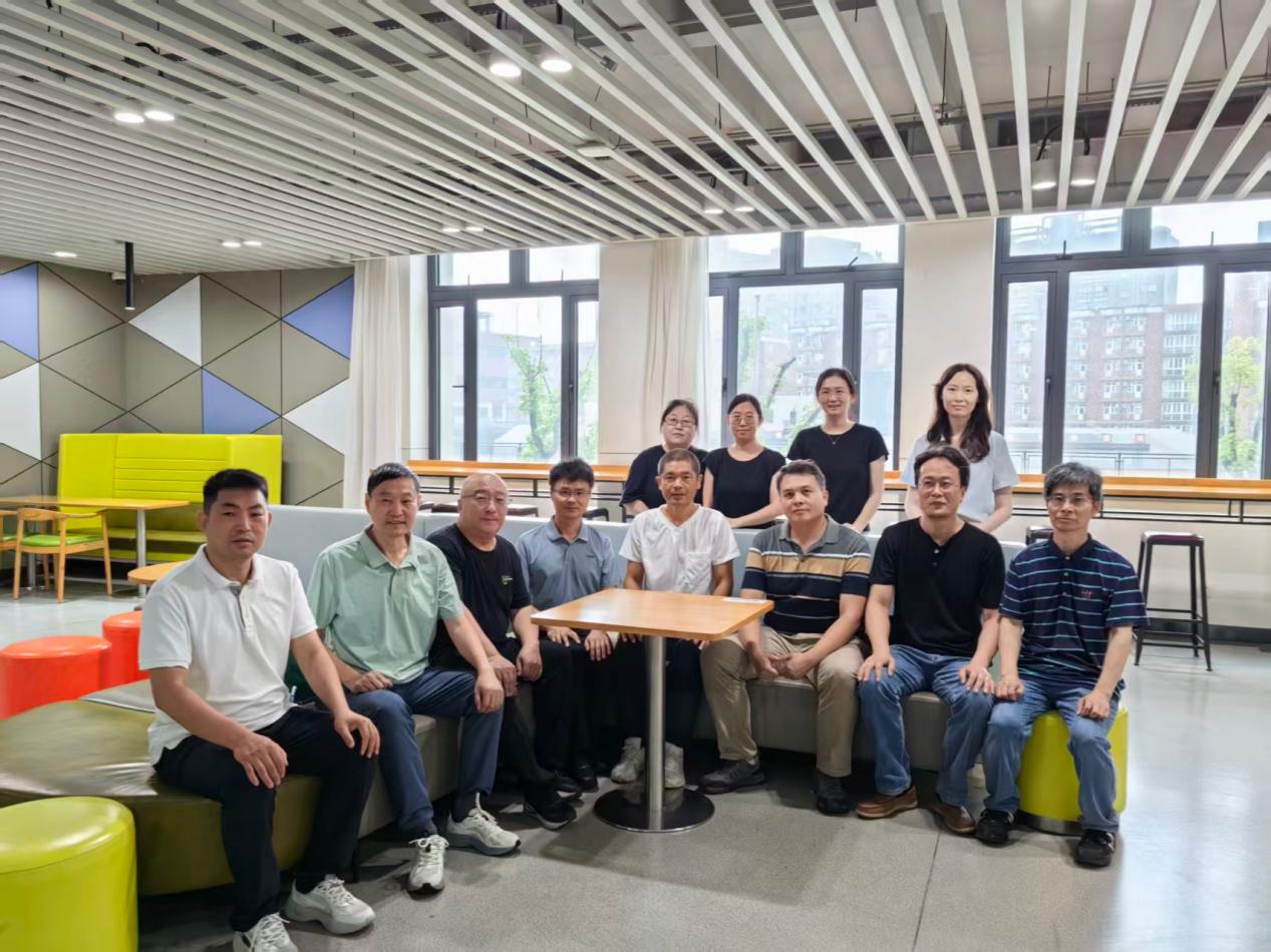
团队负责人
团队成员
蔡文杰、周雷、陈兆学、刘颖、王丽嘉、武杰、章浩伟、李丕丁、陆宏伟、沈诞、王艳、许红玉
团队简介
本团队主要从事“医学成像技术(X-ray、CT、MRI、PET、SPECT等)、智能医学信号与影像数据处理、AI 辅助疾病诊断与治疗”等方向的研究工作,以医工交叉为研究特色,是学校医疗器械学科领域的重要研究分支。近年来的研究内容包括:扩散磁共振脑微结构成像方法研究,数字化磁共振谱仪、辐射剂量模型研究;医学信号数据驱动与知识驱动融合方法,中医信号的数字化规范;人工智能辅助乳腺癌的良恶性鉴别以及分子分型自动分类研究,人工智能辅助甲状腺疾病早期诊断算法等。 团队面向临床,主攻智能影像工程领域的关键技术,致力于解决临床疾病诊疗中的医工交叉技术性问题、医学影像设备的系统工程问题等,争取建成国内外医学影像工程领域有影响力的团队。
代表性成果
[1] 2026-2029年,国家自然科学基金-面上项目,模态缺失下眼眶MRI跨模态生成和联合表征及甲状腺眼病智能诊疗研发
[2] 2025-2027,横向项目,基于人工智能的脑血管病侧支循环评估和再灌注预后预测
[3] 2025-2026,横向项目,人工智能预测股骨脆性骨折患者死亡率的模型开发
[4] 2024-2026,横向项目,乳腺癌超声图像及数据分析
[5] 2018-2023,上海市自然科学基金-面上项目,基于弥散张量成像的群体无偏脑图谱构建方法研究
2.发明专利
[1] 王丽嘉, 裴孟超, 李建奇, 聂生东, 王远军. 一种获取造影剂弛豫时间的方法[P]. 中国专利: ZL201410470363.1.
[2] 王丽嘉, 裴孟超, 董芳, 李建奇, 聂生东, 王远军. 一种利用自回归模型计算磁共振图像表观弥散系数的方法[P]. 中国专利: ZL 201410361347.9.
[3] 王丽嘉, 裴孟超, 李建奇, 王乙. 一种基于磁共振成像对穿支皮瓣血管定位与测量的方法[P]. 中国专利: ZL 201210235419.6.
[4] 王丽嘉, 裴孟超, 李建奇. 一种全自动分割量化心脏磁共振图像左心室的方法[P]. 中国专利: ZL 201110027847.5.
[5] 王丽嘉; 裴孟超; 李建奇;一种全自动分割量化心脏磁共振图像左心室的方法[P],中国专利,ZL201110027847.5.
3.学术论文
[1] Prediction Crphn’s Disease Activity Using Computered Tomography Enterography-based Radiomics and Serum Markers. Academic Radiology, 2025.
[2] DMAF-Net: deformable multi-scale adaptive fusion network for dental structure detection with panoramic radiographs. Dentomaxillofacial Radiology, 2024,53(5):296-307.
[3] MMANet: A Multi-Task Residual Network for Alzheimer’s Disease Classification and Brain Age Prediction, IRBM, 45: 100840, 2024.
[4] Multi-input dense convolutional network for classification of hepatocellular carcinoma and intrahepatic cholangiocarcinoma, Biomedical Signal Processing and Control,2023, 80: 104226, 2023.
[5] ECG classification based on guided attention mechanism. Computer Methods and Programs in Biomedicine, 2024, 257:108454.
[6] Hydrogen sulfide functions as a micro-modulator bound at the copper active site of Cu/Zn-SOD to regulate the catalytic activity of the enzyme. Cell Reports, 2023,42(7):112750.
[7] Prototype Learning Guided Hybrid Network for Breast Tumor Segmentation in DCE-MRI. IEEE Transactions on Medical Imaging, 2025, 44(1):244-258.
[8] Automated Segmentation by SCA-UNet can be Directly used for Radiomics Diagnosis of Thymic Epithelial Tumors. European Journal of Radiology, 2025, 112004.
[9] Development and verification of a novel system for computed tomography scanner model construction in Monte Carlo simulations, Nuclear Engineering and Technology , 2025, 54:4244-4252
[10] Automated pancreatic segmentation and fat fraction evaluation based on a self-supervised transfer learning network,Computers in Biology and Medicine, 2024, 170: 107989